Have you noticed that as we grow older, aging eyelids is our eyelids begin to show signs of aging—losing their firmness, appearing droopy, and making the eyes look smaller or sleepy?
Causes of Aging Eyelids: Age-related Changes in the Eyelids
Anatomical changes affecting both the skin and deeper tissues—such as thinning skin, loss of elasticity, and descent of fatty tissue, muscle, or ligaments—contribute to eyelid aging. Common aging-related eyelid issues include upper eyelid sagging, brow drooping, and increasingly prominent under-eye bags.
Learn more about common eyelid problems that develop with age and how they affect appearance and vision.
For the upper eyelids, the most common eyelid conditions are classified into three main categories:
- Dermatochalasis
- Blepharoptosis / Eyelid ptosis
- Brow ptosis
Dermatochalasis
Excess upper eyelid skin occurs due to loss of elasticity over time. In younger individuals, especially among certain ethnic groups, genetic factors may also contribute (e.g., single eyelid with fat accumulation). Elderly patients often experience concurrent upper eyelid droop (blepharoptosis) or brow ptosis, which must be distinguished clinically for appropriate treatment.
Symptoms of Dermatochalasis
Eyelid skin laxity often causes excess upper eyelid skin to droop, leading to a tired appearance, narrower double eyelids, or even the look of single eyelids. In severe cases, drooping eyelids may obstruct peripheral vision, especially while driving, and force patients to overuse forehead muscles, resulting in fatigue and permanent wrinkles. Inward-turning lashes can also irritate the cornea, causing discomfort or injury. Early recognition and treatment of drooping eyelids can help restore both vision and aesthetics.
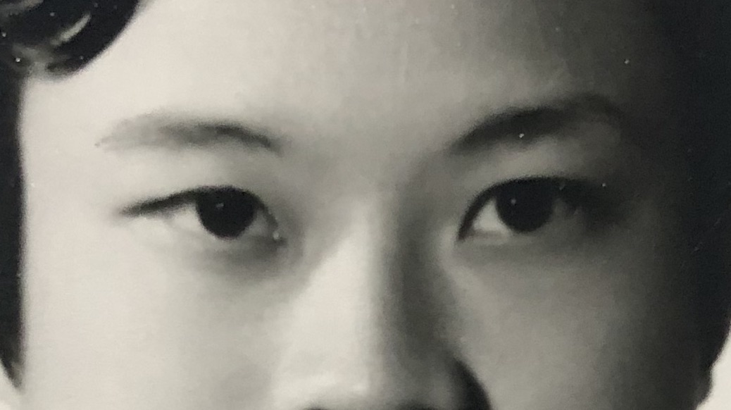
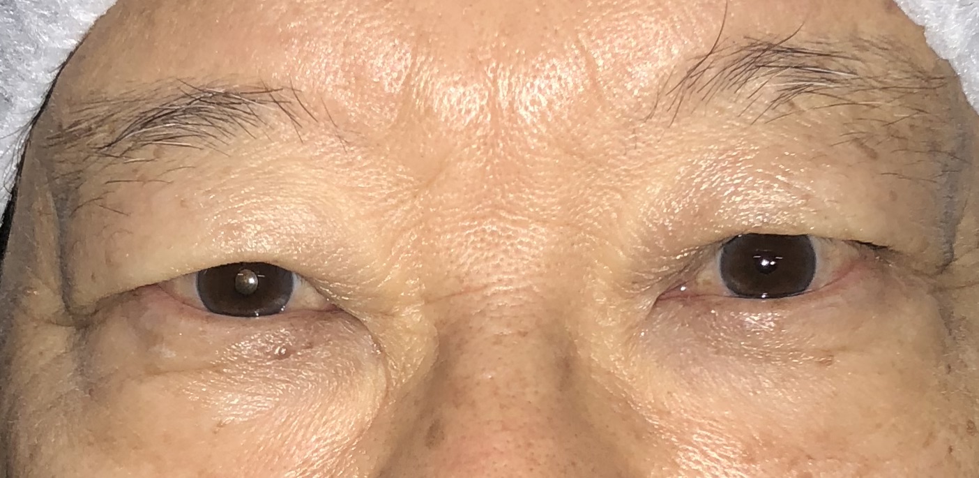
These two images demonstrate the effect of aging on the upper eyelids. By comparing photographs of the same individual at the age of 24 and 74, one can clearly observe how five decades have led to significant drooping of the upper eyelid skin, almost covering the eyelashes. In addition, the eyebrows appear elevated due to the constant use of forehead muscles to compensate and maintain clear vision
How to Treat Dermatochalasis
Droopy eyelids can be effectively corrected through upper eyelid surgery (upper blepharoplasty). The procedure involves removing the excess sagging skin of the upper eyelid. The incision is carefully placed along the natural eyelid crease (if present), allowing the scar to be well-concealed. During surgery, any excess fat that contributes to eyelid puffiness may also be removed. The incision is then closed meticulously to restore a natural and aesthetically pleasing eyelid contour.
In some cases, upper blepharoplasty can be combined with double eyelid surgery to enhance the eyelid fold. This is achieved by securing the skin to the levator muscle, creating a defined crease when the eyes are open.

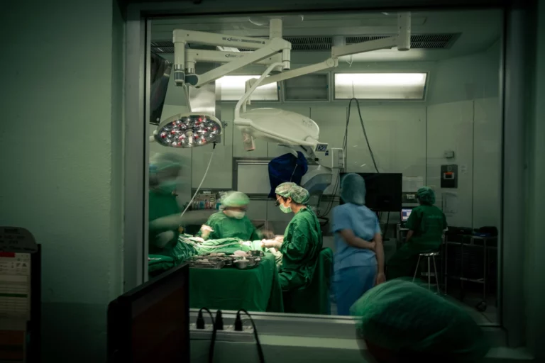
This image demonstrates upper blepharoplasty, a surgical procedure to correct sagging upper eyelids.
Blepharoptosis / Eyelid ptosis)
Ptosis (Drooping Eyelids) Ptosis refers to a condition in which the upper eyelid droops lower than its normal position. Under normal circumstances, the upper eyelid margin rests just above the upper edge of the cornea. When the eyelid droops significantly—close to or covering the pupil—it not only gives the eyes a tired or half-closed appearance but can also interfere with vision and the visual field.
A common cause of ptosis in older individuals is the weakening or detachment of the levator muscle, which is responsible for lifting the eyelid. When this occurs, surgical correction to tighten or reattach the levator muscle is often required to restore proper eyelid function and improve both vision and appearance.
Symptoms of Ptosis
When the upper eyelid droops and covers the pupil, it may cause the following symptoms:
If ptosis occurs in only one eye, it can cause noticeable asymmetry between the eyes and eyebrows. The affected side may appear to have a higher eyelid crease and a deeper eye socket, resulting in an unbalanced appearance. This is because people naturally tend to raise the eyebrow on the side with the drooping eyelid, often unconsciously, in order to improve vision.
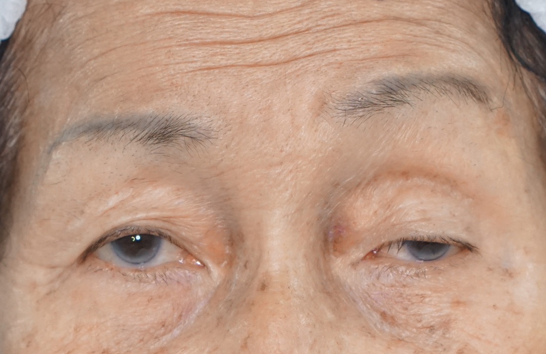
This image demonstrates left upper eyelid ptosis, where the drooping eyelid partially covers the corneal light reflex, leading to visual obstruction in the left eye. The left eyelid also appears deeper set, with the left eyebrow positioned higher than the right due to compensatory eyebrow elevation. Additionally, forehead creases are visible, caused by overactive frontalis muscle effort to lift the drooping eyelid.
When both upper eyelids droop, the eyes may appear half-closed, giving a constantly drowsy look. Both eyebrows are often elevated as the forehead muscles work harder to compensate, which can lead to forehead strain, eyebrow fatigue, and permanent forehead creases. Patients may also experience a narrowed upper visual field, reducing the ability to see widely. Some individuals unconsciously tilt their head back to improve vision, which may cause neck discomfort or pain over time.
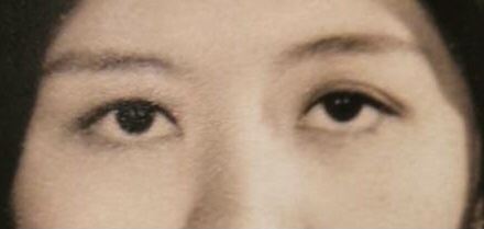
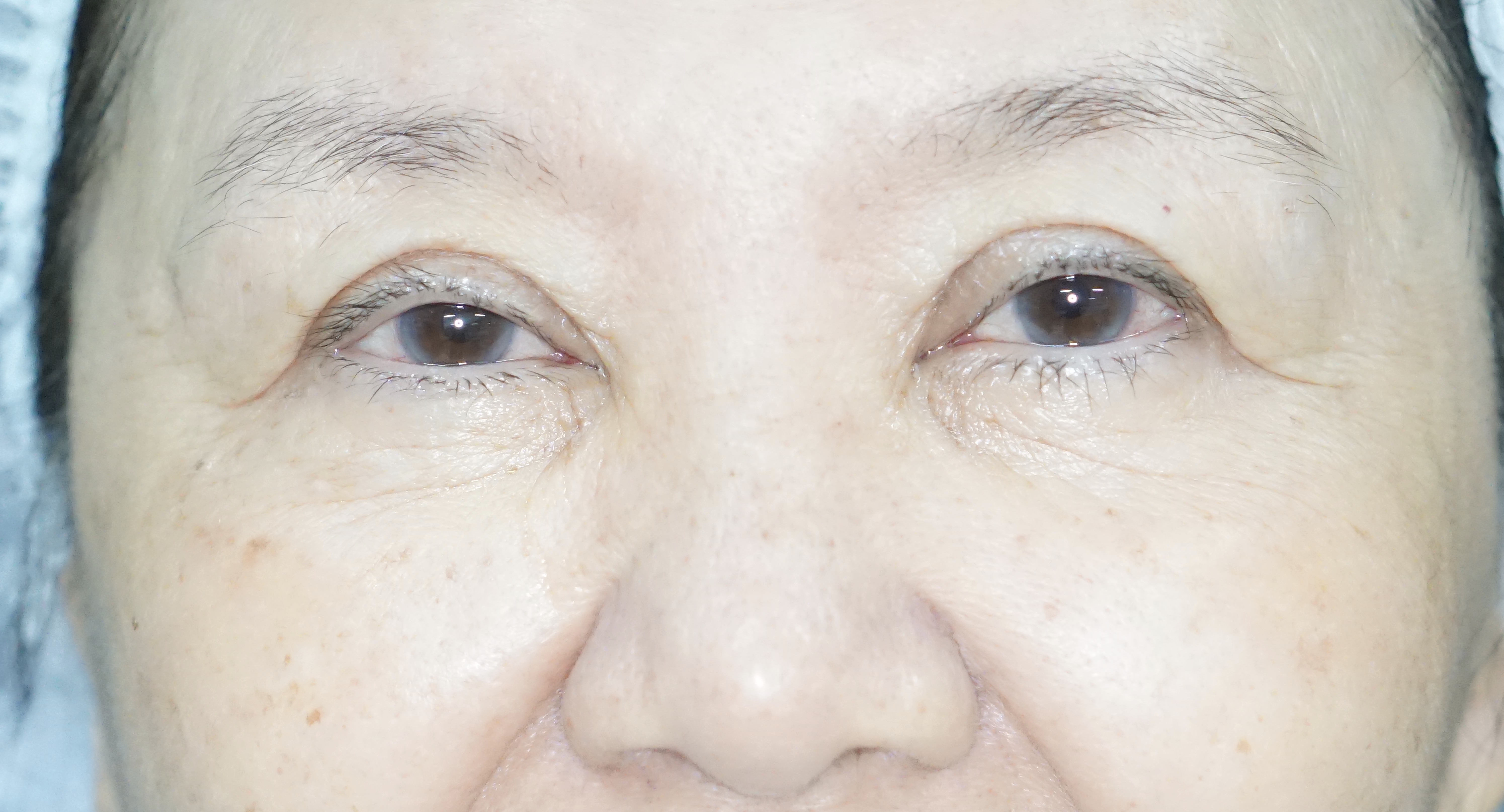
These two photographs demonstrate bilateral eyelid ptosis associated with aging, comparing the same individual at age 65 to age 25. Notice how the upper eyelid margin has descended, covering more of the cornea and reducing the margin reflex distance-1 (MRD-1)—the distance between the upper eyelid margin and the corneal light reflex. This shortening gives the eyes a droopier, more tired appearance. In addition, there is mild lateral hooding of the upper eyelid skin, further contributing to the aged look.
How Is Ptosis Treated?
Since ptosis is primarily caused by dysfunction of the muscles responsible for eyelid elevation, its correction differs from an upper blepharoplasty, which mainly involves the removal of excess skin and superficial tissue as previously described. Ptosis surgery, on the other hand, addresses the deeper layer at the level of the eyelid lifting muscles. The two main muscles responsible for elevating the upper eyelid are:
- Levator muscle
- Müller’s muscle
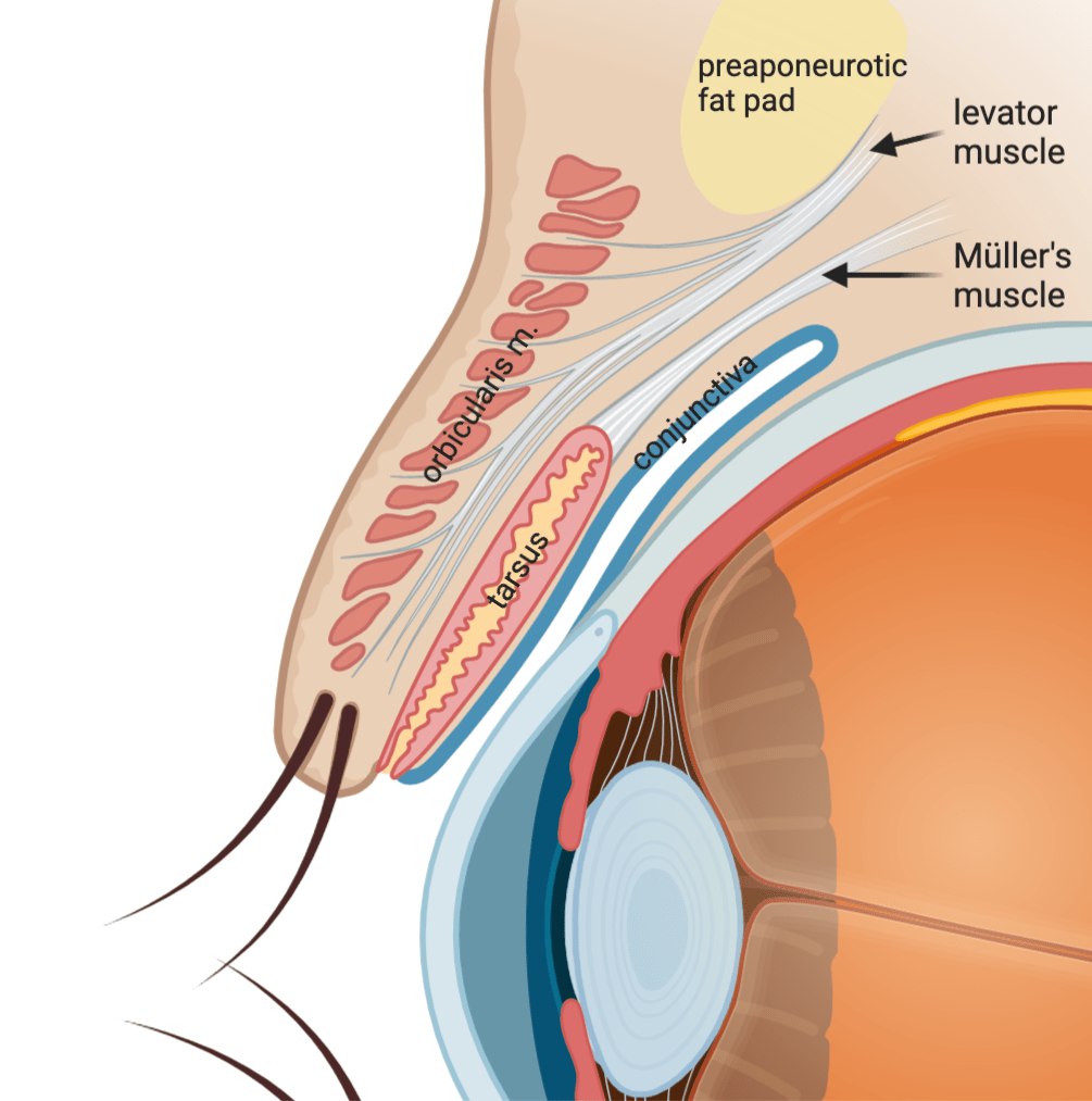
The surgical approach to correcting ptosis depends on the severity of the eyelid droop and the residual strength of the eyelid muscles. In age-related ptosis, the most common cause is the disinsertion or weakening of the levator aponeurosis, the primary muscle responsible for lifting the upper eyelid.
In cases of severe ptosis, correction is often performed using an external levator advancement. This involves creating an incision along the natural eyelid crease and reattaching or tightening the levator aponeurosis to restore proper eyelid elevation. In patients who also present with dermatochalasis (excess upper eyelid skin), the procedure can be combined with the removal of redundant skin while simultaneously tightening the levator muscle, resulting in both functional and aesthetic improvement.
In cases of mild ptosis, particularly when there is a positive response to phenylephrine testing (a medication that dilates the pupil and stimulates normal function of the Müller’s muscle), correction can be achieved with an internal eyelid approach called conjunctivomullerectomy. During this procedure, the eyelid is everted, and an incision is made through the conjunctiva to tighten the Müller’s muscle. This technique avoids any external skin incision, leaving no visible scar, while effectively improving eyelid position.
It is important to note that surgical techniques and expertise may vary among surgeons. Therefore, choosing a specialist with extensive experience is essential, as they can accurately evaluate each patient’s condition and recommend the most appropriate surgical approach tailored to individual needs.


